Table of Contents
The nose is a prominent structure between the eyes that acts as the entrance to the respiratory system and includes the olfactory organ. It provides the air we need to breathe, enables the sense of smell, filters the air, warms and moisturizes it, ventilates it, and cleanses itself of foreign particles that enter the body when inhaled.
The nose has two cavities that are separated by a cartilaginous wall called the septum. External openings are known as nostrils. The roof of the mouth and the floor of the nose are made of the palatine bone, which is usually called the hard palate. A portion of the soft palate tissue expands into the nasopharynx (the part that connects the nose to the throat) and is compressed upwards when swallowed, so it closes the nasopharynx so that food does not fit in the back of the nose.
The shape of the nasal cavity is complex. The front, inside and top of each nostril is called the nasal cavity. Behind the nasal cavity and along each outer wall there are three protrusions that generally extend from front to back. Each protrusion, called the nasal concha, or turbine, hangs from an airway. Next to and above, the upper part of the concha is the olfactory part of the nasal cavity. The rest of the cavity is the respiratory tract. The respiratory tract is covered with a moist mucous membrane with hair follicles called eyelashes, which are used to collect waste products. Mucus of membrane cells also helps trap dust particles, carbon, soot and bacteria. The sinus cavities in the bony skull are on either side of the nose.
Many things happen below the surface of the nose. Bones and cartilage under the skin make up most of the nose. Other structures inside and behind the nose help to breathe. Learning nasal anatomy can help you better understand how your nose works.
Nasal bone
The nasal bone supports the nasal bridge. The upper cartilage supports the lateral part of the nose. The lower cartilage increases its support, width and height, and helps shape the nostrils and tip of the nose.
The nasal bones are two small, symmetrical middle bones of the skull that form the bridge of the nose. The upper bouts featured two cutaways, for easier access to the higher frets, and the lower bouts featured two cutaways, for easier access to the higher frets.
Nasal skin
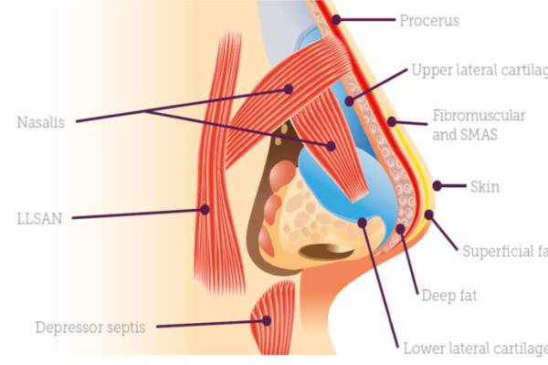
The thickness of the nasal skin varies in different parts. From the eyebrows or the distance between the eyebrows to the bridge (nose angle) the skin of the nose is thick, flexible and moveable. This thickness becomes narrower in the upper part of the nose and is placed in the thinnest position and the least flexibility because it is close to the lower bone. From the bridge to the tip of the nose, the skin is thin. The tip of the nose is covered with skin that is as thick as the upper part and has large sebaceous glands. The thickness of the skin varies but is still separated by four layers of underlying bone and cartilage – a layer of superficial fat, a layer of muscle, a layer of deep fat and a periosteum.
Nasal cavity
The nasal cavity is the hollow space behind the nose through which air passes. The anatomy of the nasal cavity is essential for both respiration and sense of smell. Did you know that 80% of taste is actually due to smell? This is why food is almost tasteless when we have a nasal congestion.
The nasal cavity is a large, air-filled space at the top and back of the nose in the middle of the face. The nasal septum divides the cavity into two parts, also called fossae. Each cavity is a continuation of one of two nostrils. The nasal cavity is the upper part of the respiratory system and provides the nasal passage for inhaled air from the nostrils into the nose and the rest of the airways./ Rhinoplasty in Iran
Nasal septum
The septum is a thin wall made of cartilage and bone that divides the inside of the nose into two parts. The nasal septum is the main supporting structure of the midline of the nose and consists of quadrangular cartilage, the vertical plane of the ethmoid bone, and the femur.
The outer fleshy end of the nasal septum is called the columella and is made up of cartilage and soft tissue. The nasal septum contains bony cartilage and vitreous cartilage. It is usually about 2 mm thick.
Mucous membranes
The mucous membrane, also known as the mucosa, is a layer of cells that surrounds organs and ducts. Made of extracellular tissue. Mucous membranes may contain or secrete mucus, a thick fluid that protects the inside of the body from contamination and pathogens such as viruses and bacteria. There are many mucous membranes in the body, such as mucous membranes in the respiratory tract, gastrointestinal tract, and genital system.
The nasal mucosa is a thin tissue that covers the nose, sinuses, and throat. It warms and moistens the breathing air. It also helps the sticky mucus to clean dust and other small particles in the air.
Nasal conchae or turbine
Turbinates also known as the nasal conchae, are the crustal networks of bones, arteries, and tissues in the nasal passage. These structures are responsible for heating, humidifying and filtering the air we breathe. There are usually three tentacles in the nose, including the upper, middle, and lower tentacles. However, sometimes there may be a fourth nasal concha (called the largest nasal concha) that is higher than the upper nasal concha.
There is a space (known as meati) between each nasal concha, each with a name that corresponds to the name of the nasal concha that is directly above the space. These spaces form the nasal passages that direct airflow through the nose.
As mentioned, the nasal tentacles are divided into three parts, upper, middle and lower.
Lower concha
The space between the floor of the nasal cavity and the lower tentacle, which is the largest airway. This duct has several features:
- The nasolacrimal duct (tear duct), which drains any secretions from the eyes, starts in the outer eye and drains into the lower tentacle.
- The apex of the nasal wall, the lower tentacle, and the bony piriform opening form the nasal valve. The nasal valve is the narrowest area in the nasal cavity and is often blocked due to septal deviation or other nasal abnormalities.
Middle concha
The nasal passage between the lower and middle horns is important in the following cases:
- Three paranasal sinuses, including the maxillary sinus, the frontal sinus, and the anterior ethmoid sinus, are drained into this space.
2. Air flows through the paranasal sinuses that make up the tone of our voice
Upper concha
This space is usually the highest nasal passage, however, sometimes there is an upper tentacle that is above the upper tentacle. The functions of this duct include the following:
- Discharge of two paranasal sinuses including sphenoid sinus and posterior ethmoid sinus.
- Like the middle tentacle, airflow through this duct (which interacts with the sinus cavities) helps to improve our acoustic properties.
- The mucous membranes of the upper tentacles (along with the upper part of the nasal septum that forms the left and right nostrils), which are covered with nerve bases, are used to aid in smell. For this reason, disturbances in the tentacles may impair the sense of smell and taste.
The upper and middle tentacles are part of the ethmoid bone, but the lower tentacles are an independent structure.
Nasal sinuses
The paranasal sinuses are a group of four air-filled spaces that surround the nasal cavity. The maxillary sinuses are located under the eyes. The frontal sinuses are located above the eye. The ethmoid sinuses are located between the eyes and the sphenoid sinuses are behind the eye. The sinuses are named after the facial bones in which they are located.
Humans have four paired nasal sinuses that are divided into subgroups that are named based on the bones in which the sinuses are located:
The maxillary sinuses, the largest paranasal sinuses, are located under the eyes, in the maxillary bones. They are innervated by the trigeminal nerve.
The frontal sinuses, above the eyes, are located in the front bone, which forms the hard part of the forehead. These sinuses are also innervated by the trigeminal nerve.
The sphenoid sinuses, which are made up of several separate air cells in the ethmoid bone between the nose and eyes. This sinus is innervated by the sphenoid nerves, which branch from the optic nerve and the trigeminal nerve.
Sphenoid sinuses located in the Sphenoid bone. These sinuses are innervated by the trigeminal nerve.
The paranasal sinuses are covered with the respiratory epithelium.